


A Case Report of Umbilical Cord Haematoma Resulting in Fresh
Still Birth in University College Hospital, Ibadan, Nigeria.
Margaret F. Ogunkuade1, Bankole A. Adewumi1, Olubukola A. Adesina1,2
1 – Department of Obstetrics and Gynaecology, University College Hospital, Ibadan, Nigeria.
2. college of Medicine, University of Ibadan, Ibadan, Nigeria.
Corresponding Author:
Dr Olubukola A. Adesina
Email: bukiadewole@gmail.com
Phone number: 08033486836
Summary
Umbilical Cord Haematoma (UCH) is a very rare but fatal obstetric complication that is poorly reported especially in Nigeria and there is the need to encourage the reporting of every case in order to expand the knowledge about this condition with the goal of reducing the associated morbidity and mortality. We report here 23year old primigravida at gestational age of 40weeks and 2days diagnosed with preeclampsia. She had an emergency lower segment caesarean section on account of fetal bradycardia. She was delivered of a fresh still born with an umbilical cord haematoma. Umbilical haematoma, though rare could be a cause of fetal bradycardia and all cases should be reported and reviewed in other to improve the global knowledge and reduce the associated fatality rates.
Introduction
Still births have remained a major cause of concern for both the obstetrician and expectant mothers. The cause of still births are many with a vast majority being unexplained. Umbilical haematoma, a form of placental cord abnormality has been reported as a rare cause of fetal still birth. Umbilical cord haematoma is said to occur when there is extravasation of blood, mainly venous, in the Warton’s jelly that covers the umbilical vessels. The umbilical cord contains umbilical vessels (two arteries and one vein) surrounded by a gelatinous stroma (Wharton’s jelly) and covered by a single layer of amnion. The arteries carry deoxygenated blood from the fetus to the placenta while the vein carries oxygenated blood from the placenta to the fetus. Umbilical cord haematoma generally occurs following the rupture of an umbilical vessel, mostly the vein with subsequent extravasation of blood into the Warton’s jelly covering the umbilical vessels1-4
We present here a 23year old primigravida at gestational age of 40 weeks and 2 days diagnosed with preeclampsia complicated by umbilical cord haematoma resulting in fresh still birth.
Case Presentation
Mrs. O.O., a booked 23year old primigravida at gestational age (GA) of 40weeks and 2days was admitted into the Labour ward complex of the University College Hospital on the 9th May, 2022 from the Antenatal Clinic of the hospital on account of preeclampsia (blood pressure (BP) of 140/90mmHg, proteinuria of 2+). She was commenced on Magnesium Sulphate (MgSO4) for seizure prophylaxis and scheduled for Induction of Labour (IOL) the following day.
She had no complaints at presentation in the labour ward. There was no labour pain, no passage of show, no drainage of fluid or bleeding per vaginum. She had no complaints of headache, visual disturbances, epigastric pain, or reduced urine output. She however affirmed to reduced perception of fetal movement occurring after her admission.
The index pregnancy was spontaneously conceived, and she booked at GA of 15weeks and 4days with normal parameters. Her antenatal period has been uneventful until this visit at 40weeks and 2days. She had a Doppler obstetric ultrasound on admission which showed a single live fetus in longitudinal lie, cephalic presentation with estimated fetal weight of 3.76kg, amniotic fluid index of 11.1cm3 and anterofundal placenta. However, the Doppler indices showed reduced end-diastolic flow signifying mild uteroplacental insufficiency, hence the scheduled IOL.
She attained menarche at 12years of age and menstruates for 3-5days in a regular cycle of 28days. Her last menstrual period (LMP) was 31st July, 2021 with her expected date of delivery (EDD) being 7th May, 2022. She had no history of menorrhagia, dysmenorrhea or dyspareunia. She had no preexisting medical co- morbidity and no prior history of surgery or blood transfusion. Her blood group was O Rh (D) Positive while her haemoglobin genotype was homozygous AA.
She was a government employee and the only wife of a self-employed farmer. Both had tertiary level of education. Both her grandmother and father were known hypertensives. She neither used tobacco nor consumed alcohol and she had no known allergies.
On examination in the labour ward, she was afebrile, not pale, anicteric, not dehydrated and she had no pedal oedema. Her blood pressure was elevated (150/90mmHg) while other vital signs were within normal limits. There were no palpable uterine contractions. The symphysiofundal height (SFH) was 38cm with a singleton fetus in longitudinal lie and cephalic presentation palpated. The Fetal heart rate was 140/min. No other organs were palpable. Pelvic examination revealed no cervical dilatation or effacement.
Her packed cell volume (PCV) was 34%, repeat urinalysis showed proteinuria of 2+ and other parameters were normal. Other investigations - Full blood count (FBC), Electrolyte, Urea and Creatinine (E/U/Cr), clotting profile, liver function test and 24hour urine protein were assayed. She was commenced on MgSO4 seizure prophylaxis using the Zuspan regimen. She had Cardiotocograph (CTG) machine applied for continuous fetal monitoring. Non - Stress test on admission was reactive. However, few hours into admission, CTG recorded bradycardia of 106-112b/m. Fetal resuscitation was commenced by keeping her in the left lateral position, administering humidified intranasal oxygen and intravenous 0.9% normal saline while written informed consent was obtained for an emergency lower segment caesarean section (EMLSCS). The interval from diagnosis to intervention was about 20 minutes as there was no significant improvement in the fetal parameter with the resuscitative measures. Preparation for EMLSCS was ensured within this period.
She had the EMLSCS and the intraoperative findings included a poorly oxygenated uterus evidenced by minimal dark coloured blood, well formed lower uterine segment, copious meconium-stained amniotic fluid, a 3.45kg female fresh still born (FSB) delivered cephalad with a tight nuchal cord. The umbilical cord was multiply twisted with a haematoma on a large portion and a gangrenous part of about 3cm length which was less than 2cm from the placenta. The estimated blood loss was 300mls.
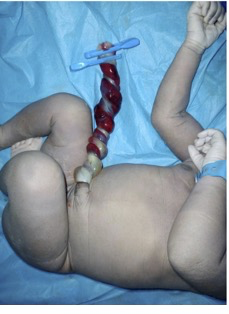
Fig. 1 – the case reported fresh still born with umbilical cord haematoma
The patient and relatives were empathised with, counselled and educated about the findings at surgery. The request for an autopsy was declined by the patient and relatives.
She had no post- operative complication. Her BP post- operative was 110-135/70-85mmHg with no usage of any antihypertensives. She was discharged from the hospital on the 4th post- operative day after counselling on breast management and contraception. She had follow-up in the clinic at 2weeks and 6weeks postpartum with normal BP and stable clinical status and subsequently discharged to the family planning clinic.
Discussion
The umbilical cord contains umbilical vessels (two arteries and one vein) surrounded by a gelatinous stroma (Wharton's jelly) and covered by a single layer of amnion. The arteries carry deoxygenated blood from the fetus to the placenta while the vein carries oxygenated blood from the placenta to the fetus. Umbilical Cord Haematoma generally occurs following the rupture of an umbilical vessel and subsequent extravasation of blood into the Warton’s jelly covering the umbilical vessels1-4. The umbilical vein is the vessel that is most times ruptured and only in about 10% of cases has arterial bleeding been noted4, 5.
It is a rare condition that is associated with still births6 with an incident rate of 1 in 5,505 to 1 in 110,000 pregnancies3. This is despite umbilical cord abnormalities being rated as the second commonest cause of still births7. The perinatal mortality rate of umbilical cord haematoma is estimated as about 50%8. The rarity of this condition may explain why no case of umbilical cord haematoma was found published in Nigeria during literature searches as at the time of writing this report.
The exact cause of umbilical cord haematoma is not known but risk factors include abnormal cord length and features (a short cord, twist, knot or prolapse), umbilical vessel aneurysm, trauma, infection, entanglement of the cord, coagulation disorder, post maturity and iatrogenic (e.g. umbilical vessel venipuncture, amniocentesis)2, 8. In the reported case above, the exact cause could not be ascertained but the nuchal cord3, twisted cord6, preeclampsia9 and postdated pregnancy5 may be contributory factors. There was no coagulation disorder in the patient and no infection diagnosed.
The diagnosis of umbilical cord haematoma is difficult during pregnancy as the case may follow a benign course, hence, the diagnosis is made postnatally on examination of the baby, umbilical cord and placenta3. This occurred in the case reported here. However, some features that can bring to suspicion about the condition include reduced perception of fetal movement, abnormal fetal heart rate tracings, ultrasound findings of notable cystic umbilical cord masses and the presence of Doppler velocimetry abnormalities1, 10. The hematomas may appear as solid masses of variable shape and size in close relation to the umbilical cord or have a variable appearance, depending on the time elapsed between the bleeding and sonographic evaluation10. While acute hematomas appear isoechoic or may be heterogeneous, chronic hematomas appear hypoechoic to anechoic on ultrasound scan10. When umbilical cord haematoma is diagnosed intrapartum, there is need for the immediate delivery of the fetus due to the high risk of fetal death associated3. In this reported case, there was no ultrasound diagnosis of the fetal haematoma though there was Doppler finding of a reduced end diastolic flow which signifies a mild uteroplacental insufficiency, hence the scheduled IOL. However, this did not establish the diagnosis of umbilical cord haematoma because of the associated finding of preeclampsia9 in the patient. Fetal bradycardia was picked in this case necessitating an emergency Caesarean delivery but unfortunately, a fresh still born was the outcome. This emphasises the need to be vigilant, and prompt in a bid to reduce the mortality rate associated with umbilical cord hematoma.
The complications of umbilical cord haematoma include severe umbilical cord compression due to large hematomas resulting in fetal anoxia and distress. The fetal distress upon the delivery of baby may result in admission into the Neonatal Intensive Care Unit (NICU), hypoxic ischaemic encephalopathy and stillbirths8. Fetal death may result from anoxia due to compression of fetal vessels by the haematoma or by the exsanguination of fetal blood1. About half of the cases of umbilical cord hematomas usually result in fetal death or still birth8. For the live deliveries, there is no need for any specialized care of the cord after birth as the haematoma should be left to fall off with the umbilical cord6.
A detailed physical examination of the placenta and cord confirms the presence of umbilical cord haematoma5. During the macroscopic examination, the umbilical cord may have abnormal appearance like the twisted dark red and bluish appearances in this case report3. Histopathological examination of the placenta, cord and still born, if available, essentially show perivascular haemorrhagic infiltration, the compressed umbilical vessels, fissures of the walls of the affected blood vessels, moderate inflammatory leukocytic infiltration of the vascular wall3,5. However, in the case reported, there was no histopathology findings as the patient and her relatives declined the request for autopsy of the still born or sending the placenta for pathology review.
Conclusion
There is poor awareness about umbilical cord haematoma especially in Nigeria as there has been no case reported in literature as at the time of this report. It is important to understand the associated risk factors, clinical manifestations and the need for expedient management. All cases of umbilical cord haematoma are encouraged to be reported in order to improve the global knowledge and reduce the associated mortality rates.
References
- Mota F, Oliveira N, Fonseca M, Mimoso G. Spontaneous umbilical cord haematoma. BMJ Case Rep. 2019;12(6).
- Cunningham FG, Leveno KJ, Bloom SL, Dashe JS, Hoffman BL, Casey BM, et al. Editors. Williams Obstetrics, 25e. New York, NY: McGraw-Hill Education; 2018.
- 3. Scutiero G, Bernardi G, Iannone P, Nappi L, Morano D, Greco P. Umbilical Cord Hematoma: A Case Report and Review of the Literature. Obstet Gynecol Int. 2018;2018:2610980.
- 4. Scutiero G, Bernardi G, Iannone P, Nappi L, Morano D, Greco P. Corrigendum to "Umbilical Cord Hematoma: A Case Report and Review of the Literature". Obstet Gynecol Int. 2018;2018:1405281.
- 5. Khatiwada P, Alsabri M, Wiredu S, Kusum V, Kiran V. Spontaneous Umbilical Cord Hematoma. Cureus. 2021;13(2):e13048.
- 6. Arora PK, Mohandas S, McAndrew S, Karody V. Spontaneous Umbilical Cord Hematoma. J Pediatr. 2017;184:233- e1.
- 7. Nappi L, Trezza F, Bufo P, Riezzo I, Turillazzi E, Borghi C, et al. Classification of stillbirths is an ongoing dilemma. Journal of perinatal medicine. 2016;44(7):837-43.
- 8. Abraham A, Rathore S, Gupta M, Benjamin SJ. Umbilical Cord Haematoma Causing Still Birth- A Case Report. J Clin Diagn Res. 2015;9(12):QD01-2.
- 9. Olaya-C M, Salcedo-Betancourt J, Galvis SH, Ortiz AM, Gutierrez S, Bernal JE. Umbilical cord and preeclampsia. Journal of Neonatal-Perinatal Medicine. 2016;9:49-57.
- 10. Sherer DM, Al-Haddad S, Cheng R, Dalloul M. Current Perspectives of Prenatal Sonography of Umbilical Cord Morphology. Int J Womens Health. 2021;13:939-71.